

Otosclerosis
Otosclerosis is a condition in which the bones in the middle ear become fixed and immobile, leading to hearing loss (usually one-sided).Treatment for Otosclerosis
There are three options in the management of otosclerosis.
No Treatment
The first involves no treatment and coping with the degree of hearing loss that is present and following the hearing with serial audiograms.
Hearing Aids
Hearing aids have been found to be useful in otosclerosis provided the clarity is still preserved. These devices amplify all sound they receive. This is one of their drawbacks. The brain has a remarkable ability to focus in on sounds we want to hear and dampen sounds that are undesirable. Hearing aids, with all their amplification, prevent the brain's ability to focus.
SURGERY
Stapedectomy is a microsurgical procedure that I perform under general anesthesia. The procedure takes approximately one hour and is typically done in an ambulatory surgery center. Patients go home the same day. Typically, the procedure is done on Thursday or Friday and patients are back to work by Monday or Tuesday.
The entire operation is performed through the ear canal with an operating microscope. An incision is made in the ear canal skin and the eardrum is elevated. A small nerve called the chorda tympani that transmits taste information from the front 2/3 of the tongue frequently has to be moved to allow adequate visualization of the stapes. (Temporary taste disturbance is not uncommon after this operation and usually resolves in a few months.) The ossicles are then examined and the joint between the incus and stapes is severed. A laser is used to divide a tendon, which holds the stapes in place. The stapes is then fractured and removed. A laser is then used to vaporize a small area in the footplate of the stapes. The stapes bone is a triangulated bone with the base of the triangle facing the inner ear. This base is called the footplate. Fragments of the footplate are then gently picked away from the oval window. This defect in the footplate is then covered with a piece of vein harvested from the hand. A tiny prosthesis is inserted over the vein graft and attached to the incus.
There are two basic techniques used in otosclerosis surgery:
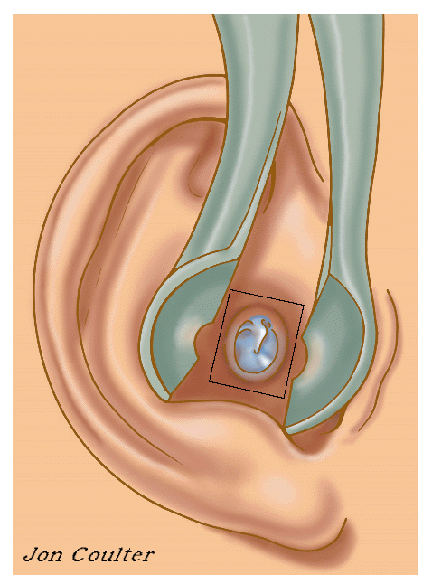
Step 1: For orientation the ear, ear canal and eardrum are shown. The square is enlarged in the next drawing.
Step 2: An incision is made in the ear canal skin adjacent to the eardrum.
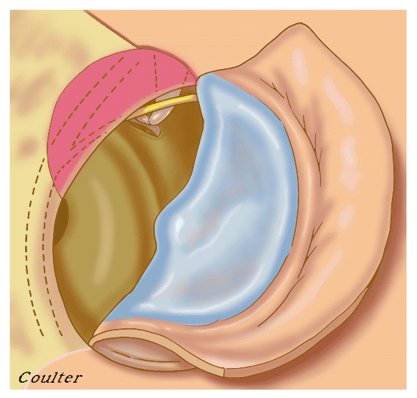
Step 3: The ear canal skin along with a portion of the eardrum is elevated. The shaded area represents the area of bone that must be removed to allow adequate visualization of the stapes bone.
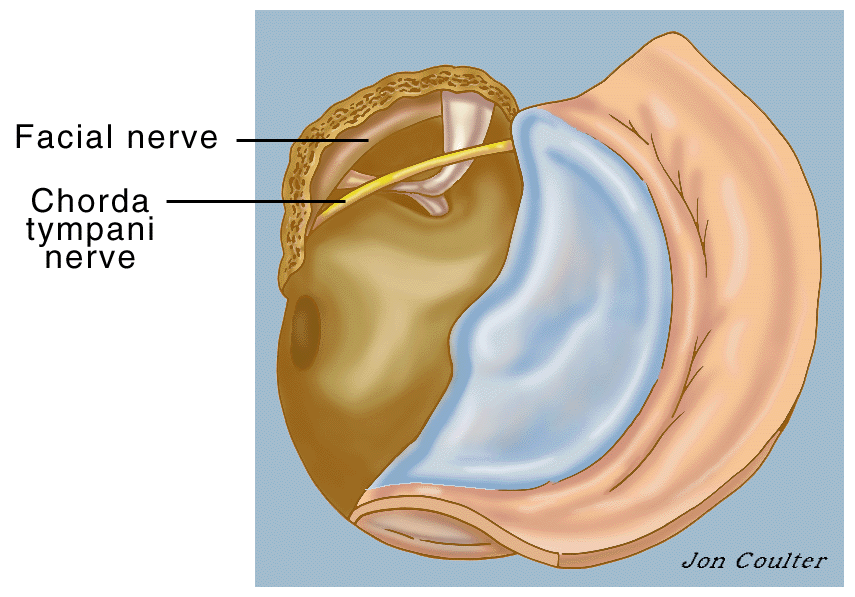
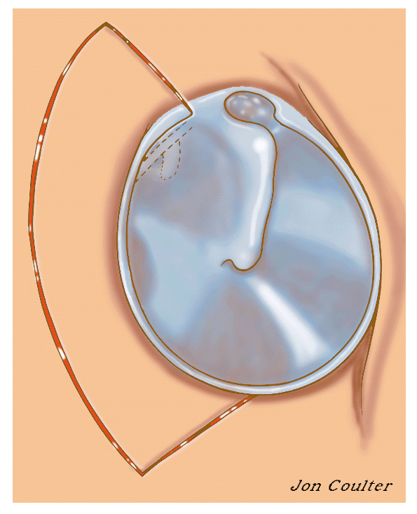
Step 4: The chorda tympani nerve that transmits information about taste from the tongue frequently has to be moved around. A segment of the facial nerve shown is usually covered with bone. The incus and stapes bones are seen better on the next enlarged view.
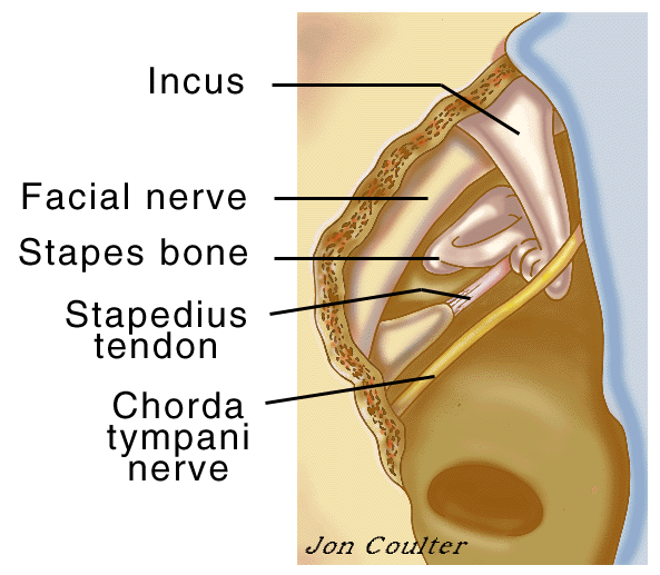
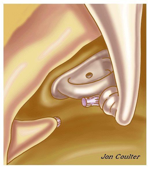
Step 5: The incus, stapes, stapedius tendon, chorda tympani nerve and facial nerve are shown.
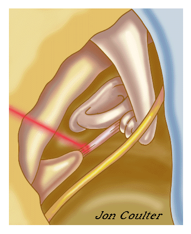
Step 6: A laser is used to divide the stapedius tendon.
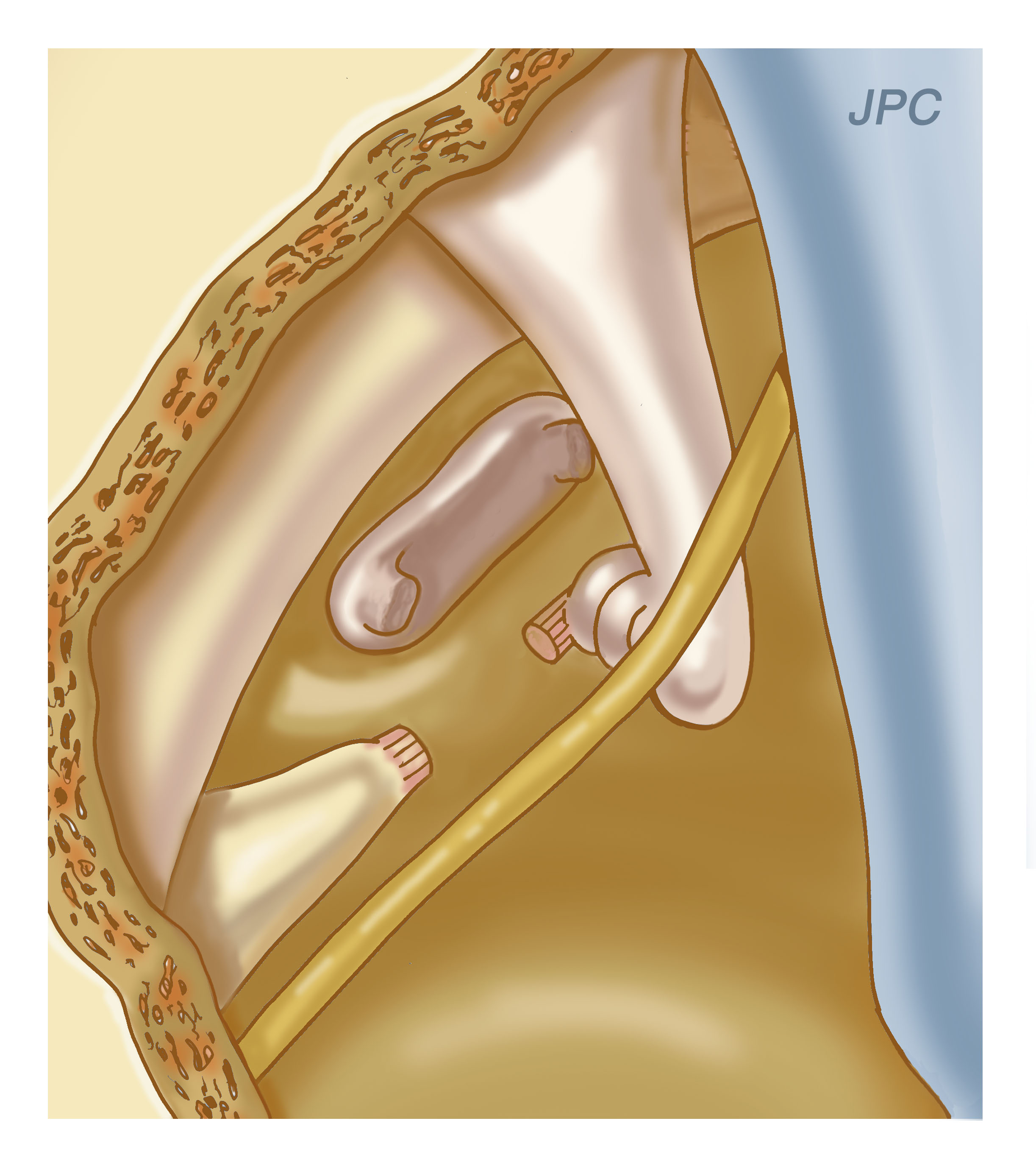
Step 7: One of the arches of the stapes bone is divided and the joint between the incus and stapes bones is separated. The outer part of the stapes bone is removed. For illustrative purposes, the chorda tympani nerve is not shown on successive drawings.
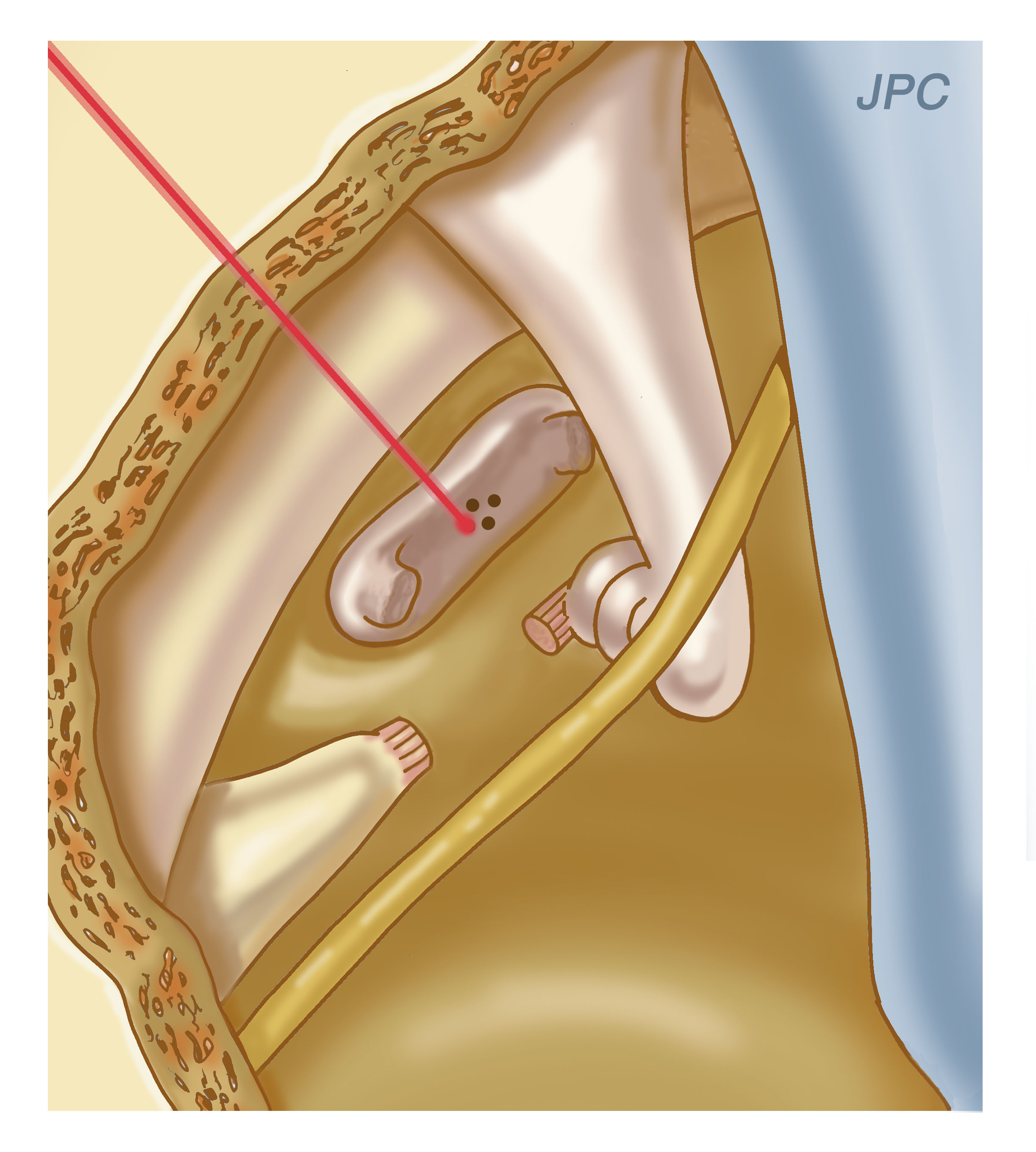
Step 8: A laser is used to make a hole in the footplate of the stapes bone.
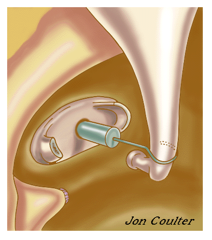
Step 9: A prosthesis is inserted and attached to the incus bone.
Small Fenestra Technique
The small fenestra technique involves making a small hole in the footplate of the stapes and inserting the prosthesis through that hole. Please click on the diagram on the left below. This will show the steps of the operation.
Step 1: For orientation, the ear, ear canal, and eardrum are shown. The square is enlarged in the next drawing.
Step 2: An incision is made in the ear canal skin adjacent to the eardrum.
Step 3: The ear canal skin along with a portion of the eardrum is elevated. The shaded area represents the area of bone that must be removed to allow adequate visualization of the stapes bone.
Step 4: The chorda tympani nerve that transmits information about taste from the tongue frequently has to be moved around. A segment of the facial nerve shown is usually covered with bone. The incus and stapes bones are seen better on the next enlarged view.
Step 5: The incus, stapes, stapedius tendon, chorda tympani nerve, and facial nerve are shown.
Step 6: The stapes tendon is divided, and a small hole is made in the central part of the footplate. The joint between the incus and stapes bones is disrupted.

Step 7: The stapes bone is removed.

Step 8: A graft is placed over the oval window. A prosthesis is placed over the graft and attached to the incus bone.
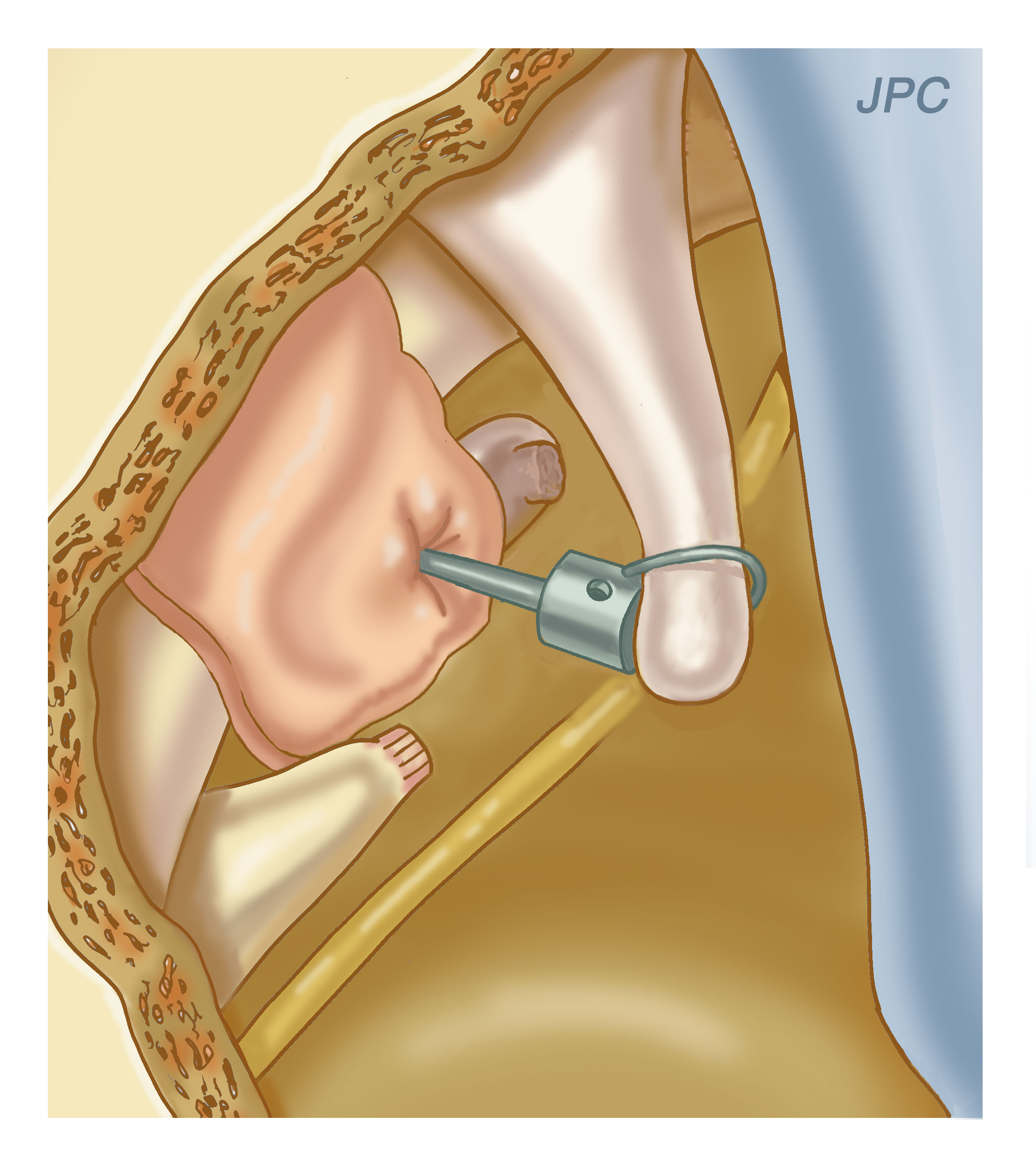
Step 9: A prosthesis is placed over the graft and attached to the incus bone.
Wide Fenestra Technique
The wide fenestra technique involves removing as much of the footplate that will easily come out. A graft is then placed over the oval window (the opening to the inner ear from the middle ear) and the prosthesis is then placed over the graft. This is the technique that I prefer. Please click on the diagram on the right below. This will show the steps of the operation.
It must be remembered that not all otolaryngologists (ear, nose, and throat physicians) perform this operation on a regular basis. Most physicians who perform this type of operation have successfully completed a fellowship in neurotology and limit their practice to ear surgery.
We are not in the business of training medical students, residents or fellows. Doctors-in-training will not be operating on you. I perform all these procedures from the beginning to the end.
No one should scare you from having a stapedectomy operation for fear of facial paralysis or hearing loss. It is the most successful procedure for hearing loss that I perform. Many health care professionals are more interested in selling you a hearing aid every 5 years than doing an insurance-covered procedure that can last 20 years.
About Dr. Prasad

Dr. Sanjay Prasad MD FACS is a board certified physician and surgeon with over twenty years of sub-specialty experience in Otology, Neurotology, advanced head and neck oncologic surgery, and cranial base surgery. He is chief surgeon and founder of the private practice, Metropolitan NeuroEar Group, located in the metropolitan Washington D.C. area.
Find us on Facebook
Follow us on Twitter
- Otosclerosis tweeted Our website has launched! http://t.co/gnZyv4zIOctober 27, 2011 at 11:19am
- Otosclerosis tweeted Visit our brand new educational website, http://t.co/0nTHIrH1, for more information about hearing loss related to otosclerosis!October 27, 2011 at 11:18am
- Otosclerosis tweeted Have you been diagnosed with otosclerosis? Need information on what it is? Want access to others with the same condition? Follow us!January 07, 2011 at 12:25pm